45 microscope images with labels
Compound Microscope: Definition, Diagram, Parts, Uses ... Compound microscope is a type of optical microscope that is used for obtaining a high-resolution image. There are more than two lenses in a compound microscope. Learn about the working principle, parts and uses of a compound microscope along with a labeled diagram here. Sharper microscope images wanted: labels need not apply ... Sharper microscope images wanted: labels need not apply 9 posts JournalBot. Ars Legatus Legionis et Subscriptor. Registered: Apr 5, 2005. Posts: 101520. Posted: Thu Oct 30, 2008 2:06 pm ...
LAS X Industry Microscope software for Industry | Products ... Create a single sharp image by capturing a stack of images at different focus positions and combining them automatically into an Extended Depth of Focus (EDOF) image. LAS X Extended Depth of Field: Create sharp 2D images from several partially in-focus images. In connection with the 3D Surface Viewer, creation of 3D images is also possible.

Microscope images with labels
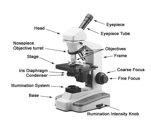
Parts of a Simple Microscope - Labeled (with diagrams ... image 5: A modern simple microscope with the different parts labeled. image source: laboratoryinfo.com The optical parts of a simple microscope are centered on the specimen - lighting, and magnification. Microscope Imaging Station. Gallery. - Exploratorium Elodea leaf cells with structures labeled Chloroplasts and mitochondria move within Elodea leaf cells; nuclei are also visible as clear, fried-egg-shaped structures. Elodea are common freshwater aquarium plants. More images in this category: Elodea leaf cells Spirogyra Elodea leaf cells with structures labeled Spirogyra, structures labeled Parts of the Microscope with Labeling (also Free Printouts ... Microscopes are specially created to magnify the image of the subject being studied. This exercise is created to be used in homes and schools. the microscope layout, including the blank and answered versions are available as pdf downloads. Click to Download : Label the Parts of the Microscope (A4) PDF print version.
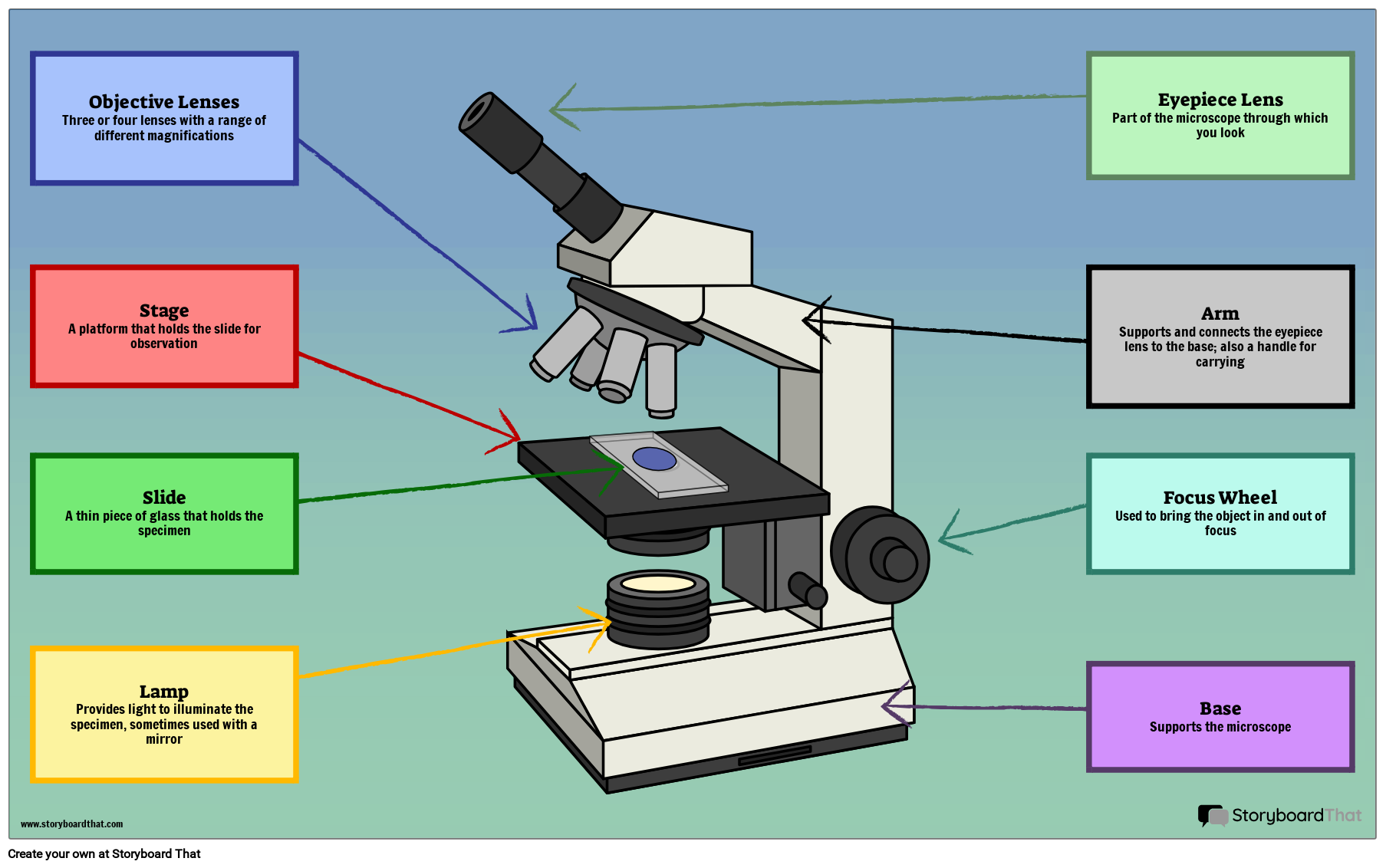
Microscope images with labels. › en › microscopeFluorescence Resonance Energy Transfer (FRET ... - Olympus In most cases, however, the normal diffraction-limited fluorescence microscope resolution is insufficient to determine whether an interaction between biomolecules actually takes place. Fluorescence resonance energy transfer is a process by which radiationless transfer of energy occurs from an excited state fluorophore to a second chromophore in ... Parathyroid Gland Histology with Microscope Slide Image ... Parathyroid Gland Histology with Microscope Slide Image and Labeled Diagram 09/12/2021 07/12/2021 by anatomylearner There are two or more pairs of parathyroid glands located on the posterior surface of the thyroid gland. You will find two main types of cells (chief and oxyphils) in the parathyroid gland histology slide. Microscope Parts and Functions With Labeled Diagram and ... First, the purpose of a microscope is to magnify a small object or to magnify the fine details of a larger object in order to examine minute specimens that cannot be seen by the naked eye. Here are the important compound microscope parts... Eyepiece: The lens the viewer looks through to see the specimen. Microscope Types (with labeled diagrams) and Functions A compound microscope: Is used to view samples that are not visible to the naked eye. Uses two types of lenses - Objective and ocular lenses. Has a higher level of magnification - Typically up to 2000x. Is used in hospitals and forensic labs by scientists, biologists and researchers to study micro organisms. Compound microscope labeled diagram.
PDF Label parts of the Microscope Label parts of the Microscope: . Created Date: 20150715115425Z ZEISS Axiocam Microscope Cameras for Science and Research For easy documentation, some digital cameras can record images either completely without a computer, or through a connected PC, laptop or iPad running the ZEISS Labscope software. All our microscope cameras are fully supported in our ZEN software environment, offering fast live image display and easy-to-use user interface. Microscope Images Labeled | Virtual Anatomy Lab VAL Microscope Images Labeled | Virtual Anatomy Lab VAL Microscope Labeling Game - PurposeGames.com About this Quiz. This is an online quiz called Microscope Labeling Game. There is a printable worksheet available for download here so you can take the quiz with pen and paper. This quiz has tags. Click on the tags below to find other quizzes on the same subject. Science.
Electron microscope - Wikipedia An electron microscope is a microscope that uses a beam of accelerated electrons as a source of illumination. As the wavelength of an electron can be up to 100,000 times shorter than that of visible light photons, electron microscopes have a higher resolving power than light microscopes and can reveal the structure of smaller objects.. Electron microscopes use shaped magnetic … 125,082 Microscope Photos - Free & Royalty-Free Stock ... Browse 125,082 professional microscope stock photos available royalty-free. Details of medical laboratory, scientist hands using microscope for chemistry test samples. Details of medical laboratory, scientist hands using microscope for. Scientist hands with microscope, examining samples and liquid. Medical research with technical equipment. DinoCapture 2.0: Microscope Imaging Software - Dino-Lite Dino-Lite USB microscope cameras include DinoCapture 2.0, the powerful yet easy to use microscope imaging software for Windows. DinoCapture is a professional microscope imaging software that was made for users of all levels, including basic features from image viewing and capture, measurement with calibration, to advanced features such as geotags and edge … Compound Microscope with labels Stock Vector | Adobe Stock Compound Microscope with labels Stock Vector | Adobe Stock. Get 10 free Adobe Stock images.
Flares into Darkness: Insect faces
What is Electron Microscopy? - UMASS Medical School Because of its great depth of focus, a scanning electron microscope is the EM analog of a stereo light microscope. It provides detailed images of the surfaces of cells and whole organisms that are not possible by TEM. It can also be used for particle counting and size determination, and for process control. It is termed a scanning electron microscope because the image is formed by …
31 Picture Of Microscope With Label - Labels Database 2020
Label the microscope - Science Learning Hub All microscopes share features in common. In this interactive, you can label the different parts of a microscope. Use this with the Microscope parts activity to help students identify and label the main parts of a microscope and then describe their functions.. Drag and drop the text labels onto the microscope diagram. If you want to redo an answer, click on the box and the answer will go back ...
Print Microbiology Lab 2 (Microscopy, Gram stain, intro to enterotube II) flashcards | Easy ...
Microscope, Microscope Parts, Labeled Diagram, and Functions Microscope, Microscope Parts, Labeled Diagram, and Functions What is Microscope? A microscope is a laboratory instrument used to examine objects that are too small to be seen by the naked eye. It is derived from Ancient Greek words and composed of mikrós, "small" and skopeîn,"to look" or "see".

Beyond the Human Eye: A tiny aquatic worm that clones itself
300+ Free Microscope & Laboratory Images - Pixabay Upload 399 Free images of Microscope Related Images: laboratory science bacteria research scientist lab biology chemistry medical Find your perfect microscope image. Free pictures to download and use in your next project. 399 Free images of Microscope / 4‹ ›

labels of a compound microscope microscope boxed - Top Label Maker
Microscope Drawing And Label at PaintingValley.com ... Are you looking for the best images of Microscope Drawing And Label? Here you are! We collected 33+ Microscope Drawing And Label paintings in our online museum of paintings - PaintingValley.com. ADVERTISEMENT LIMITED OFFER: Get 10 free Shutterstock images - PICK10FREE label microscope diagram compound parts light labeling functions microscopic

Microscope Picture To Label - Micropedia
Compound Microscope Parts - Labeled Diagram and their ... The eyepiece (or ocular lens) is the lens part at the top of a microscope that the viewer looks through. The standard eyepiece has a magnification of 10x. You may exchange with an optional eyepiece ranging from 5x - 30x. [In this figure] The structure inside an eyepiece. The current design of the eyepiece is no longer a single convex lens.

Stacey Kalkowski's Art Journal: Pollen Spores and Asteroids
Microscope Labeled Pictures, Images and Stock Photos Browse 48 microscope labeled stock photos and images available, or start a new search to explore more stock photos and images. Newest results Fluorescent Imaging immunofluorescence of cancer cells growing... Plant Tissue Systems vector illustration. Labeled biology... Microscope diagram vector illustration. Labeled zoom instrument...

Microscopy Flipped Home Learning - Lessons - Tes Teach
Microscopic Images Of Tissues Flashcards Flashcards by ... This set of image-based flashcards gives medical students the Microscopic Images of Tissues. Learn the terms, keywords, vocabulary, and much more about Microscopic Images of Tissues with our flashcards quizzes. Attempt and answer these flashcards quizzes easily and have a smooth experience with it. Cards In This Set
1.1 Labelling Microscope - Labelled diagram
medcell.med.yale.edu › histology › urinary_system_labHistology - Yale University Answer: The kidneys would appear the same under the light microscope, but the foot processes of the podocytes would be missing in the EM of the minimal change kidney. Patients with this disease have edema because they can no longer repel proteins from entering the urine, and there is a loss of albumin from the blood into the urine, which is ...
Microscope Labeling
Microscope Objective Lens | Products | Leica Microsystems The objective lens is a critical part of the microscope optics. The microscope objective is positioned near the sample, specimen, or object being observed. It has a very important role in imaging, as it forms the first magnified image of the sample. The numerical aperture (NA) of the objective indicates its ability to gather light and largely determines the microscope’s resolution, the ...
Labelling A Microscope - Labelled diagram
PDF Parts of a Microscope Printables - Homeschool Creations Label the parts of the microscope. You can use the word bank below to fill in the blanks or cut and paste the words at the bottom. Microscope Created by Jolanthe @ HomeschoolCreations.net eyepiece head objective lenses arm focusing knob base illuminator stage stage clips nosepiece.

Skin (Integumentary System)
Amazon.com: AmScope LED-144W-ZK White Adjustable 144 LED ... This interference prevents the light from sitting square with microscope body. (See attached images)It's a good light with nice dimming feature, but since I can't mount it securely or orient it the way it was intended to be used, I am returned it.AmScope LED-56S-ZK on the other hand has the same brightness, reinforced screw holes, doesn't have ...

Exploration of the Human Spinal Cord
ZEISS Axioscope 5 - Smart Microscope for Lab Routine and ... In the past, documenting samples with multiple fluorescent labels in your routine lab could be time consuming. To get best image quality, you needed to manually switch filters, adjust illumination intensities and exposure times and to snap each single channel image. For four different channels, this could sum up to 15 steps and clicks. With Smart Microscopy, this is a …

Onion Cell, 400X | Sue Bachus | Flickr
Labeling the Parts of the Microscope | Microscope World ... Labeling the Parts of the Microscope This activity has been designed for use in homes and schools. Each microscope layout (both blank and the version with answers) are available as PDF downloads. You can view a more in-depth review of each part of the microscope here. Download the Label the Parts of the Microscope PDF printable version here.

Microscope labeling
› WAI › EMIndex of Dr.Jastrow's electron microscopic atlas Table D leads to images of electron microscopes or protocols for tissue preparation. Table E leads to the overview pages with the images of this atlas which are used in the histology course of the University of Mainz, Germany. From table F you can call up the Vocabulary of microscopic anatomy which explains some terms in German and Englisch.
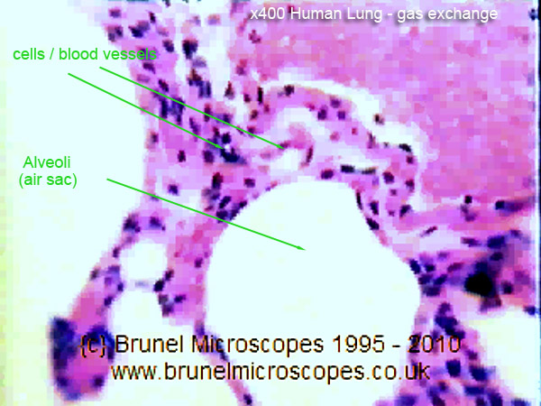
Human Cells - Part II an overview for light microscopists - Lungs
Label-free prediction of three-dimensional fluorescence ... All cell types were imaged for up to 2.5 h on a Zeiss spinning disk microscope with ZEN Blue 2.3 software and with a 1.25-NA, 100x objective (Zeiss C-Apochromat 100x/1.25 W Corr), with up to four, 16-bit data channels per image: transmitted light (either bright-field or DIC), cell membrane labeled with CellMask, DNA labeled with Hoechst, and ...
Label a microscope - Teaching resources
Microscope Labeling Practice Quiz About this Quiz. This is an online quiz called Microscope Labeling Practice. There is a printable worksheet available for download here so you can take the quiz with pen and paper.
Post a Comment for "45 microscope images with labels"