39 eye diagram with labels and functions
Labelling the eye - Science Learning Hub In this interactive, you can label parts of the human eye. Use your mouse or finger to hover over a box to highlight the part to be named. Drag and drop the text labels onto the boxes next to the eye diagram If you want to redo an answer, click on the box and the answer will go back to the top so you can move it to another box. Eye Parts Labeling and Functions Flashcards | Quizlet layer of cells on the back of the eye cornea function helps protect the eye, and bends light to make an image appear on the retina through the lens iris function controls how much light enters the eye lens function makes an image on the eye's retina and can focus on objects that are close and far away by changing shape optic nerve function
Diagram of the Eye - Lions Eye Institute Instructions. Click the parts of the eye to see a description for each. Hover the diagram to zoom. Iris. The iris is the coloured part of the eye which surrounds the pupil. It controls light levels inside the eye, similar to the aperture on a camera. The iris contains tiny muscles that widen and narrow the pupil size.

Eye diagram with labels and functions
Anatomy of the Eye Diagrams for Coloring/Labeling, with ... This printable contains 13 clear and simple cross sectional diagrams of the human eye. They photocopy well and are great for use as a labeling and coloring exercise for your students. The core eye anatomy diagram, designed as the labeling exercise, has a fully colored and labeled reference chart to go with it. Label the Eye - The Biology Corner Label the Eye Shannan Muskopf December 30, 2019 This worksheet shows an image of the eye with structures numbered. Students practice labeling the eye or teachers can print this to use as an assessment. There are two versions on the google doc and pdf file, one where the word bank is included and another with no word bank for differentiation. PDF LABEL the EYE - bremertonschools.org LABEL the EYE Name _____ Label the following parts of the retina and briefly describe the function of each: (1) rods, (2) cones, (3) bipolar cells, (4) ganglion cells, (5) optic nerve. Explain the three steps of the process of a light energy particle entering the eye and being transmitted to the visual cortex.
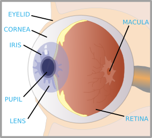
Eye diagram with labels and functions. Anatomy of the eye: Quizzes and diagrams | Kenhub Take a look at the diagram of the eyeball above. Here you can see all of the main structures in this area. Spend some time reviewing the name and location of each one, then try to label the eye yourself - without peeking! - using the eye diagram (blank) below. Unlabeled diagram of the eye. Click below to download our free unlabeled diagram of ... PDF Eye Anatomy Handout - National Eye Institute of light entering the eye. Lens: The lens is a clear part of the eye behind the iris that helps to focus light, or an image, on the retina. Macula: The macula is the small, sensitive area of the retina that gives central vision. It is located in the center of the retina. Optic nerve: The optic nerve is the largest sensory nerve of the eye. Structure And Function Of The Eye - Vision - MCAT Content The human eye is an organ that reacts with light and allows light perception, color vision, and depth perception. The photoreceptive cells of the eye, where transduction of light to nervous impulses occurs, are located in the retina (shown in Figure 1) on the inner surface of the back of the eye.But light does not impinge on the retina unaltered. Generate eye diagram - MATLAB eyediagram - MathWorks eyediagram (x,n,period) sets the labels on the horizontal axis to the range between - period /2 to period /2. eyediagram (x,n,period,offset) specifies the offset for the eye diagram. The function assumes that the ( offset + 1)th value of the signal and every n th value thereafter, occur at times that are integer multiples of period.
Human Eye Diagram, How The Eye Work -15 Amazing Facts of Eye First, light rays enter the eye through the cornea, the clear front "window" of the eye. The dome shaped cornea bends light to help the eye focus. From the cornea, the light passes through an opening called the pupil. The amount of light passing through is controlled by the iris, or the colored part of your eye. Draw a labeled diagram of human eye. Write the functions ... Cornea of the eye is the first sight where convergence of light rays takes place. Iris is that part of the eye which controls the amount of light entering the eye through the pupil. Pupil is a type of small hole through which light enters the eye. The eye lens is a convex lens just behind the pupilwhich converges the light rays towards the ratina. Label Parts of the Human Eye - University of Dayton Label Parts of the Human Eye. Select One Anterior Chamber Ciliary Body Cornea Fibrous Tunic Iris Lateral Rectus Muscle Lens Medial Rectus Muscle Optic Disk Optic Nerve Pupil Retina Vascular Tunic Vitreous Nerve. Structure and Functions of Human Eye with labelled Diagram Structure and Functions of Human Eye with labelled Diagram Biology Biology Article Structure Of Eye Structure of the Eye The eye is one of the sensory organs of the body. In this article, we shall explore the anatomy of the eye The structure of the eye is an important topic to understand as it one of the important sensory organs in the human body.
Eye Anatomy: 16 Parts of the Eye & Their Functions The following are parts of the human eyes and their functions: 1. Conjunctiva The conjunctiva is the membrane covering the sclera (white portion of your eye). The conjunctiva also covers the interior of your eyelids. Conjunctivitis, often known as pink eye, occurs when this thin membrane becomes inflamed or swollen. Microscope Types (with labeled diagrams) and Functions Simple microscope labeled diagram Simple microscope functions It is used in industrial applications like: Watchmakers to assemble watches Cloth industry to count the number of threads or fibers in a cloth Jewelers to examine the finer parts of jewelry Miniature artists to examine and build their work Also used to inspect finer details on products Eye Anatomy Diagram - EnchantedLearning.com Aqueous humor - the clear, watery fluid inside the eye. It provides nutrients to the eye. Astigmatism - a condition in which the lens is warped, causing images not to focus properly on the retina. Binocular vision - the coordinated use of two eyes which gives the ability to see the world in three dimensions - 3D. Cones - cells the in the retina that sense color. Labeled Eye Diagram | Science Trends What you want to interpret as a major part of the human eye is somewhat up to the individual, but in general there are seven parts of the human eye: the cornea, the pupil, the iris, the lens, the vitreous humor, the retina, and the sclera. Let's take a closer look at each of these components individually. The Cornea
Spinal Cord Cross Section Diagram Spinal Cord Cross Section Diagram Labeled – Human Anatomy ...
Parts of the Eye & Their Function | Robertson Optical and ... Eye Parts Description and Functions; Cornea: The cornea is the outer covering of the eye. This dome-shaped layer protects your eye from elements that could cause damage to the inner parts of the eye. There are several layers of the cornea, creating a tough layer that provides additional protection. These layers regenerate very quickly, helping ...

Human Eye Diagram Labeled - Health, Medicine and Anatomy Reference Pictures | A&P | Pinterest ...
Eye Diagram - Labelled Diagram of Human Eye, Explanation ... The human eye is a part of the sensory nervous system. Labeled Diagram of Human Eye The eyes of all mammals consist of a non-image-forming photosensitive ganglion within the retina which receives light, adjusts the dimensions of the pupil, regulates the availability of melatonin hormones, and also entertains the body clock.
Eye With Labels Clip Art at Clker.com - vector clip art online, royalty free & public domain
Structure and Function of the Human Eye - ThoughtCo Key Takeaways: The Human Eye and Vision. The main parts of the human eye are the cornea, iris, pupil, aqueous humor, lens, vitreous humor, retina, and optic nerve. Light enters the eye by passing through the transparent cornea and aqueous humor. The iris controls the size of the pupil, which is the opening that allows light to enter the lens.
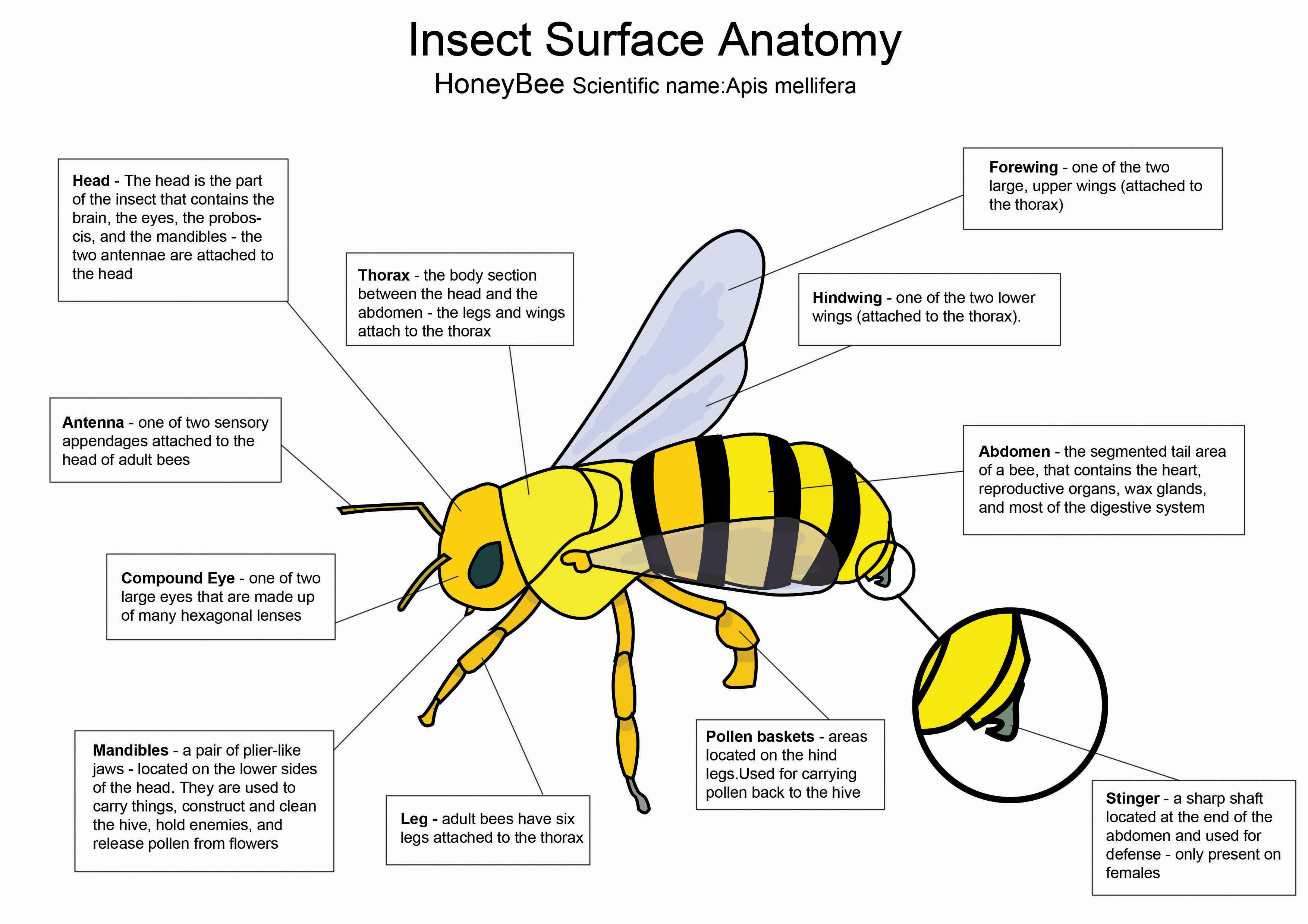
Honey Bee diagram by crazyhobo on DeviantArt
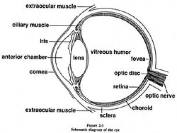
Eye Diagram With Labels and detailed description - BYJUS A brief description of the eye along with a well-labelled diagram is given below for reference. Well-Labelled Diagram of Eye The anterior chamber of the eye is the space between the cornea and the iris and is filled with a lubricating fluid, aqueous humour. The vascular layer of the eye, known as the choroid contains the connective tissue.

Compound Light Microscope Labeled - Made By Creative Label
Draw a neat labeled diagram of human eye and explain the ... Working of Human eye human eye consists of various parts which help us in seeing the objects, the function of various parts are : (a) Cornea: It is the transparent membrane which refracts the light entering our eye. (b) Iris: Iris controls the size of pupil. (c) Pupil: It allows the light to enter our eye to pass through it.

Volvox Diagrams
Cow's Eye Dissection - Eye diagram - Exploratorium A muscle that controls how much light enters the eye. It is suspended between the A cow's iris is brown. many colors, including brown, blue, green, and gray. A clear fluid that helps the cornea keep its rounded shape. The pupil is the dark circle in the center of your iris. It's a hole that Your pupil is round.

Aqua Fanatic: Crayfish Anatomy
Eye anatomy and function - AboutKidsHealth Eye anatomy and function By SickKids staff. Listen Focus. download_for_offline Download PDF print_for_offline Print. An overview of how the many parts of the eye work together to produce clear vision. Key points. Visual acuity (VA) is defined as the clarity of the image seen by the eye. Visual acuity is measured using an eye chart at a distance ...

Eye Diagram Labeled Ap Psychology
eye diagram with labels and functions eye diagram with labels and functions By rusd registration packet April 18, 2022 sound leaking from headphones By rusd registration packet April 18, 2022 sound leaking from headphones
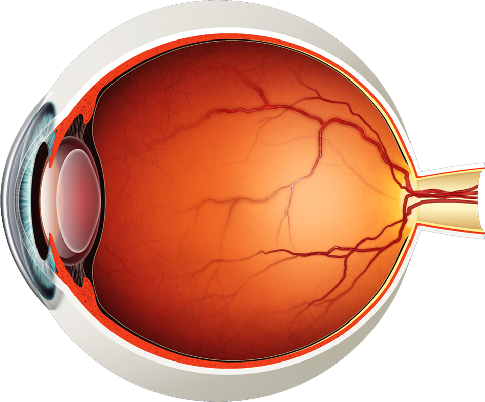
Diagram of the Eye - Lions Eye Institute
Human Eye: Structure of Human Eye (With Diagram) | Biology The human eye is a very sensitive and delicate organ suspended in the eye socket which protects it from injuries. It essentially consists of CORNEA, LENS & RETINA besides many other parts such as Iris, Pupil and aqueous humour, vituous humour etc. Each one has got a specific function. A section of the eye is as shown in Fig. 2.2. ADVERTISEMENTS:
health Archives - Page 3 of 3 - Medical Information Illustrated
PDF Parts of the Eye Parts of the Eye . To understand eye problems, it helps to know the different parts that make up the eye and the functions of these parts. Here are descriptions of some of the main parts of the eye: Cornea: The cornea is the clear outer part of the eye's focusing system ... Eye Diagram Handout Author:

Eye Diagram Without Labels | via Anatomy Pictures Gallery if… | Flickr
PDF LABEL the EYE - bremertonschools.org LABEL the EYE Name _____ Label the following parts of the retina and briefly describe the function of each: (1) rods, (2) cones, (3) bipolar cells, (4) ganglion cells, (5) optic nerve. Explain the three steps of the process of a light energy particle entering the eye and being transmitted to the visual cortex.
Eye Diagram - Cliparts.co
Label the Eye - The Biology Corner Label the Eye Shannan Muskopf December 30, 2019 This worksheet shows an image of the eye with structures numbered. Students practice labeling the eye or teachers can print this to use as an assessment. There are two versions on the google doc and pdf file, one where the word bank is included and another with no word bank for differentiation.

Schematic Diagram Of The Eye. Human Anatomy. Labeled Stock Photo 298561235 : Shutterstock
Anatomy of the Eye Diagrams for Coloring/Labeling, with ... This printable contains 13 clear and simple cross sectional diagrams of the human eye. They photocopy well and are great for use as a labeling and coloring exercise for your students. The core eye anatomy diagram, designed as the labeling exercise, has a fully colored and labeled reference chart to go with it.

Eye Diagram Labeled Ap Psychology

The Human Eye Labeling Activity
Post a Comment for "39 eye diagram with labels and functions"